Human life is unthinkable without oxygen. As trivial as this statement may sound, the processes that help transport oxygen from the ambient air into a wide variety of body cells are complex (de Marees, 2003). Due to the distance between the body's cells and the surrounding air, the human body needs special and complex transport systems, in the form of the respiratory system, cardiovascular system and the blood transport medium, to ensure an adequate supply of oxygen. Many parts of the body are involved in breathing, such as...
Breathing – Anatomy:
Upper respiratory tract in the head area:
-
Nasal cavity
-
Pharynx
-
Larynx
Lower respiratory tract in the trunk area:
-
Windpipe (trachea)
-
Tracheal branches (bronchi)
-
Pulmonary alveoli (alveoli)
Nasal cavity:
The two nasal cavities are divided into two parts by the nasal septum and separated from the oral cavity by the palate. Inside, the surface is lined with mucous membranes and covered with ciliated hairs (Fuchs & Reiß, 1990).
Respiratory functions of the nasal cavity:
-
Warming of the breathing air to up to 35-37°C through the mucous membranes, which are heavily supplied with blood
-
Humidification of the breathing air to prevent the following respiratory structures from drying out
-
Cleaning the air we breathe, through the mucous membranes and cilia, of dust and other small foreign bodies
Pharynx:
The pharynx is an approximately 10-15 cm long muscular, tube-like structure that is lined with mucous membranes and connects our mouth and nose with the food and trachea (de Marees, 2003). The pharynx also opens into the larynx, which has its own functioning.
Larynx:
The larynx adjoining the pharynx consists of several cartilages (thyroid cartilage, cricoid cartilage, arytenoid cartilage x 2), which, together with the bony tongue skeleton, form the larynx skeleton (Fuchs, 1995).
Respiratory function of the larynx:
-
Passage for breathing air as it connects the upper and lower respiratory tract
-
Protects the lower respiratory tract through a protective reflex (cough)
Windpipe (Trachea):
This is a 10-15 cm long tube-like structure that lies in front of the esophagus. Up to 20 horseshoe-shaped cartilage braces stiffen the wall of the trachea, which divides into the two main bronchi at the level of the fourth thoracic vertebra (de Marees, 2003).
Bronchi and alveoli:
The two main bronchi open into the two lungs on the right and left. There they divide into ever smaller branches (bronchioles). At the terminal bronchi there are ducts that have small, thin, shell-like bulges (alveoli or alveoli). These approximately 300 million alveoli are surrounded by a tightly knit network of pulmonary capillaries that are responsible for gas exchange (Levine / Stray-Gundersen, 1997).
Functional principle of the lungs and gas exchange:
In order to carry out gas exchange quickly and sufficiently and to reach all tissue structures, two major mechanisms are at work in humans:
-
The rapid transport of gas through the movement of gases or liquids.
This refers to the transport of gases through the respiratory tract. Through the bellows-like system (lungs, thorax, respiratory muscles).
-
rapid transport of gases through the vascular system through the heart, which acts like a valve pump (de Marees, 2003).
-
The relatively slow gas exchange through diffusion between alveoli and capillaries or capillaries and cells. So that the gas exchange can be carried out as quickly and in sufficient quantities at these points, the diffusion distances are kept short (1/1000mm and less) and the exchange areas are large (lung capillary surface = approx. 100m² / muscle capillary surface = approx. 600m²)(Levine / Stray -Gundersen, 1997).
The ribcage (thorax) consists of the sternum, ribs and thoracic spine. The ribs can be moved through joint connections between the ribs and the spine. This causes the interior of the thorax to become larger or smaller (de Marees, 2003).
The active process of inhalation (inspiration) occurs through the contraction of the diaphragm and the external intercostal muscles. These contractions cause the interior of the throat to expand and thus create a negative pressure, which results in the lungs being filled with air. The air we breathe in usually has an oxygen content of 20.9%. In the alveoli, these oxygen particles diffuse into the blood and reach the cells in which they are metabolized. The breakdown products, such as carbon dioxide, are released back into the blood by the capillaries on the cells and transported to the lungs. The passive process of exhalation occurs when the muscles relax. This is accompanied by a reduction in the size of the interior of the thorax and the escape of gases from the lungs (de Marees, 2003).
When breathing deeply, as during physical exertion, in addition to the elements mentioned above, the so-called auxiliary respiratory muscles (cervical muscles, chest muscles, anterior serratus muscles) are also involved in inspiration. This is the case as soon as the body requires a ventilated air volume of 50 l/min or more (de Marees, 2003).
During expiration, the abdominal muscles and the inner intercostal muscles help to shrink the interior of the thorax and thus ensure sufficient gas exchange (Fuchs, 1995).
Contact
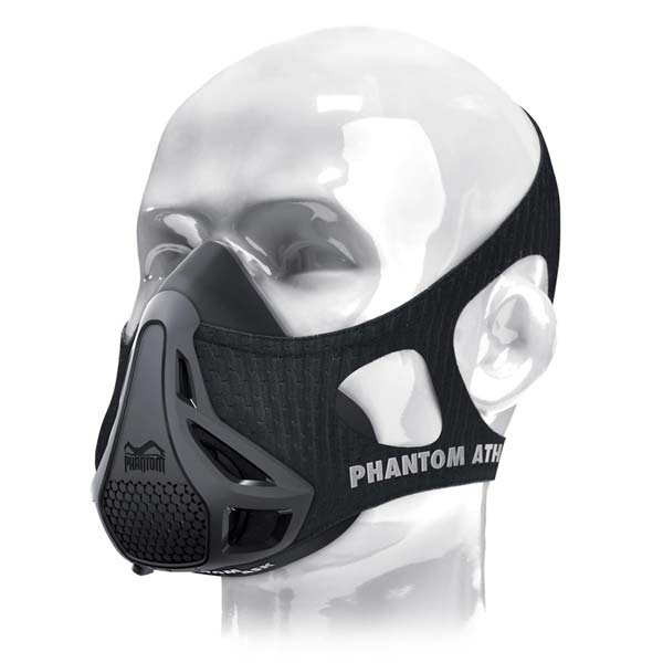
Do you have any questions about the Phantom training mask? You can contact us at any time here:
-
email: info@phantom-athletics.com
-
Tel.: +43 (0) 1 325 22 58
-
Facebook: www.facebook.com/PhAthletics
-
Instagram: phantom
-
Snapchat: phantom .athl
-
WhatsApp: +43 676 3965188
–> we are happy to help!


